Endometriosis

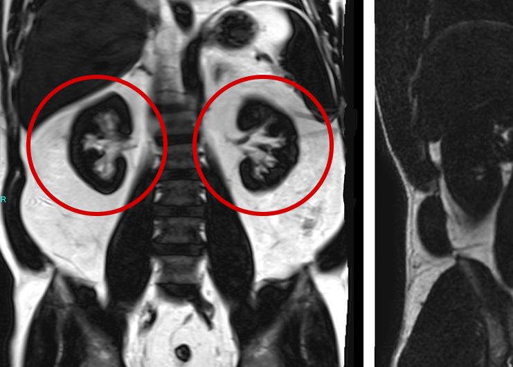
Suspected left ovarian endometrioma highlighted on a T1-weighted fat-suppressed MR image. The intensely bright signal on this sequence is characteristic of hemorrhagic material, commonly found within endometriomas. These findings reflect repeated menstrual cycle-related bleeding within the lesion, without the shedding typical of normal endometrium located in the uterus. This comprehensive approach helps accurately identify and differentiate endometriomas from other types of lesions.
Case overview
This case study focuses on endometriosis, a chronic condition affecting 10% of women of childbearing age worldwide. Often undiagnosed for years, endometriosis can cause pain, inflammation, and even infertility. Whole body MRIs can be helpful for early detection of this condition, especially if presenting symptoms are unusual or if the disease exists outside the pelvis.
Patient History
A healthy 38-year-old woman with a prior history of back pain after the birth of her first child came to Prenuvo seeking a proactive WB-MRI.
Findings
As shown in Figure 1, the Prenvuo scan showed evidence of endometriosis in the left ovary and the perineum.


Left perineal hemorrhagic lesion, which may represent an additional endometrioma
Left perineal hemorrhagic lesion, which may represent an additional endometrioma
Follow-up care
How the Prenuvo scan impacted patient care:
- Endometriosis is a chronic condition where tissue similar to the uterine lining (endometrium) grows outside of it. This can cause pain, bleeding, inflammation, and infertility, affecting about 10% of childbearing age women worldwide.
- Unfortunately, endometriosis is often missed and studies have shown an average delay of 6.7 years between the onset of symptoms (often first occurring in adolescence) and diagnosis. In this case, WB-MRI proved very useful in finding and locating endometriomas; this method can help assist healthcare providers to better manage the condition while relieving symptoms, providing an improved quality of life with less pain.
- Providers often use pelvic ultrasound or dedicated MRI to spot endometriomas and see how far the disease has spread; WB-MRI can be especially helpful for early detection, especially if presenting symptoms are unusual or if the disease exists outside the pelvis.
References
- Zondervan KT, Becker CM, Missmer SA. Endometriosis. N Engl J Med. 2020;382:1244–56. doi:10.1056/NEJMra1810764.
- Parasar P, Ozcan P, Terry KL. Endometriosis: epidemiology, diagnosis and clinical management. Curr Obstet Gynecol Rep. 2017;6(1):34-41. doi:10.1007/s13669-017-0187-1.
Other case studies
The Visceral Truth: How Advanced Imaging Detected Severe Internal Fat in an Asymptomatic Man

Subdural Hematoma
