

MRI technology represents one of the most significant breakthroughs in medical diagnostics of the 20th century. Its origins can be traced back to a fundamental discovery in quantum physics that would ultimately transform healthcare.
In 1946, physicists Felix Bloch and Edward Purcell independently discovered nuclear magnetic resonance (NMR), a phenomenon that would lay the groundwork for magnetic resonance imaging. This groundbreaking work, which earned them the Nobel Prize in Physics in 1952, was initially a pure scientific exploration with no immediate medical applications.
The journey from a theoretical physics concept to a life-saving medical tool was anything but straightforward. Early researchers recognized the potential of nuclear magnetic resonance, but the translation into medical imaging seemed like a distant dream. It took nearly three decades of persistent research, technological innovations, and interdisciplinary collaboration to bring MRI from a laboratory curiosity to a clinical reality.
The MRI pioneers
In the 1970s, three key figures made groundbreaking contributions that transformed NMR into the MRI we know today:
Raymond Damadian, an American physician and inventor, made a crucial discovery in 1971. He demonstrated that nuclear magnetic relaxation times of tissues and tumors differ, laying the foundation for using MRI as a diagnostic tool. This finding led him to propose the concept of whole body NMR scanning for cancer detection. In 1977, Damadian and his team produced the first full-body human MRI scan using a machine they called "Indomitable.”
Paul Lauterbur, a chemist, introduced the concept of gradient magnetic fields in 1973. This innovation was essential for creating two-dimensional images and marked a significant step towards practical MRI technology. Lauterbur's work on "image formation by induced local interaction" allowed for spatial localization of the MRI signal.
Peter Mansfield, a British physicist, developed mathematical techniques that enabled the rapid conversion of radio frequency signals into clear images. His work was key in improving the speed and efficiency of image acquisition.
How does MRI work? A deep dive into the science
At its core, MRI is a sophisticated blend of physics, chemistry, and biology. The technology harnesses the unique properties of hydrogen atoms—the most abundant element in the human body—and their behavior in magnetic fields. Here's a deeper dive into it:
1. Quantum mechanics meets biology: Every hydrogen atom contains a proton that spins, creating a tiny magnetic moment. In the human body, these hydrogen atoms are primarily found in water and fat molecules.
2. Magnetic field alignment: When a patient enters an MRI machine, the powerful magnetic field causes these hydrogen protons to align, like tiny compass needles pointing in the same direction.
3. Radio frequency pulses: Short bursts of radio waves temporarily knock these protons out of alignment. When the radio waves stop, the protons realign, releasing energy in the form of radio signals.
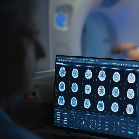
4. Signal detection and image creation: Sophisticated sensors capture these signals, which are then processed by complex algorithms to create detailed cross-sectional images of the body's internal structures.
The evolution of MRI development
1970s: Conceptual breakthroughs
- Raymond Damadian demonstrates that nuclear magnetic relaxation times of tissues and tumors differ. This discovery suggested that MRI could potentially be used to detect cancer.
- Paul Lauterbur and Peter Mansfield develop techniques for creating two-dimensional and three-dimensional magnetic resonance images. This made it possible to pinpoint where signals were coming from, allowing doctors to create detailed "slices" of the body.
- The first rudimentary MRI images are produced in low resolution.
1980s: Clinical emergence
- The first commercial MRI scanners became available.
- Gradient technology improvements dramatically enhance image resolution.
- Medical professionals start recognizing MRI's potential for non-invasive diagnostics.
1990s: Functional and specialized imaging
- Functional MRI (fMRI) was introduced, allowing visualization of brain activity.
- Cardiac and neurological imaging become increasingly sophisticated.
- Contrast agents are developed to improve imaging specificity.
2000s: Technological acceleration
- High-field strength magnets (3T and above) become more common.
- Faster scanning techniques reduce patient examination times.
- Advanced computational techniques improve image processing.
2010s and beyond: AI and precision medicine
- Machine learning and artificial intelligence begin integrating with MRI technology.
- Personalized imaging protocols become possible.
- Predictive diagnostic capabilities emerge.
Related: 1T vs. 3T: What’s the difference?
The Prenuvo difference: Industry-leading diagnostic imaging
While traditional MRI technologies have been transformative, they often fell short in several key areas:
- Long scanning times
- Limited whole body scanning capabilities
- Reactive rather than preventative approach
Prenuvo represents the culmination of decades of technological evolution. By integrating advanced AI algorithms, cutting-edge magnetic resonance technologies, and a patient-centric approach, Prenuvo has reimagined medical imaging:
- Ultra-Fast Scanning: Reduced examination times without compromising image quality
- Comprehensive Whole Body Screening: Detecting potential issues across multiple organ systems
- AI-Enhanced Diagnostics: Actively investigating AI for even better quantification and accuracy, longitudinal tracking over routine scans, and medical research breakthroughs
- Patient Comfort: Designing experiences that reduce anxiety and improve accessibility
Related: This is why everyone is getting whole body MRIs
Why does this matter now?
The evolution of MRI technology is more than just a story—it’s a call to action. Prenuvo represents the future of diagnostics, offering unparalleled early detection that could save lives. Why settle for outdated methods when you can access cutting-edge technology today?
To learn more about the benefits of a Prenuvo whole body MRI, book a call with a member of our team.