Once reserved for specific medical diagnoses, particularly for high-risk patients, and to evaluate known conditions, MRI scans such as Prenuvo’s Whole Body Scan are available as out-of-pocket, elective services for consumers in North America and globally.
Below, we breakdown the technology and clinical principles that make Prenuvo’s whole body MRI powerful for the potential of early detection.
Why whole body MRI
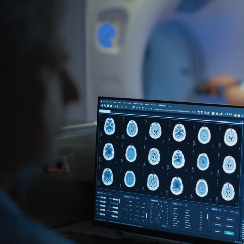
Prenuvo’s Whole Body Scans use magnetic resonance imaging (MRI), an imaging modality that leverages powerful magnetic fields to produce detailed internal images of the body. Whole body MRI can help detect a wide range of health conditions, including certain cancers, cardiovascular issues like aneurysms, neurological conditions, and musculoskeletal problems. Prenuvo’s MRI scan provides a comprehensive overview of multiple organs and tissues in a single session in less than one hour, and does not require the use of ionizing radiation or contrast injections.
Prenuvo’s MRIs offer multiparametric capabilities: MRI combines multiple “sequences,” each of which is “tuned” to highlight different tissue properties (like fluid content, fat, or cellular density), allowing radiologists to see subtle differences between normal and abnormal tissues. The more sequences (or "tissue weightings"), the better from a multiparametric MRI perspective, as they can increase sensitivity/specificity to help detect and characterize wider ranges of pathology. This tissue composition contrast capability is what Prenuvo’s MRI excels at, giving our radiologists the ability to change what is weighted, and sequences can include fluid-weighted, fat-weighted, restricted-diffusion weighted, fluid-suppressed, fat-suppressed, and others.
Prenuvo’s whole body screenings make no use of injected gadolinium-based contrast, which minimizes both invasiveness and the potential risk of accumulation from repeated uses over the long term. And unlike CT scans, Prenuvo’s MRI scans use no ionizing radiation, so they can be safely repeated over time for proactive screening without exposing patients to the cumulative radiation dose that CT delivers.
Prenuvo’s MRIs offer reliability, efficiency, and consistency
Like any advanced imaging, whole body MRI is a specialized technique that needs to be carefully optimized to provide reliable, efficient, and consistent results. Without careful thought put into the configuration of MRI hardware, image acquisition parameters, diagnostic radiology approach, and standardization of reporting of radiologic interpretations when performing screening whole body MRI — let alone emphasizing the patient experience itself to ensure it is as comfortable as possible — clinical utility of the screening whole body MRI approach can be eroded.
Prenuvo has made significant investments in our clinics, machinery, computer equipment, diagnostic software, clinical staff, radiologists, and patient comfort to ensure high and consistent levels of interpretative accuracy, reliability, and patient experience across our many locations.
What sets Prenuvo apart from other MRI screenings?
There are many things that make the Prenuvo clinical experience special. The following “characteristics” are true differentiators between us and other approaches to MRI screening.
Prenuvo optimizes voxel size

What is a voxel? It’s basically a 3D pixel. When we take images of a patient in an MRI machine, we acquire successive “slices” that we join up to form a complete, three-dimensional representation of each patient at the specific moment of the scan. Just as a 4K TV provides better image quality than a 1080p TV because it contains more pixels, an MRI acquired with more voxels—while preserving signal-to-noise-optimized parameters—allows radiologists to maximize spatial resolution, interpretative accuracy, and clinical usefulness.
Almost all MRIs are capable of taking images with voxels as small as 1mm. So you might ask, why wouldn’t a screening radiology practice take images with the smallest possible resolution? Without getting into the physics of MRI too deeply, we “construct” images of patients by listening for very faint signals given off by their bodies as we shift the magnetic field in the machine. With smaller voxel sizes, there is less signal to pick up on, which can lead to noisier images. There are basically two ways to overcome this: listen for longer to capture more signal from the smaller voxel size (which adds time to the scan), or use advanced, dedicated equipment like Prenuvo’s and place coils on the patient’s body during the scan to enhance signal pick up.
Additionally, Prenuvo’s use of tissue weighting helps drive the overall sensitivity and specificity of whole body MRI in the screening context. Because each tissue weighting is essentially an incremental acquisition sequence, the number of tissue weightings directly influences the time of the scan. The equipment and protocols used by Prenuvo enable us to acquire whole body images at diagnostic levels with more voxels per unit of time than routine MRI equipment not specifically specialized for performing screening whole body MRI. Our “time versus tissue weight” curve is similarly different to what is available at non-specialized centers. In other words, during the same amount of time spent undergoing a whole body MRI scan at Prenuvo, the radiologist receives a far broader spectrum of tissue-weighted sequences—each with high image resolution, optimized signal-to-noise characteristics, and excellent overall quality—while also covering substantially more anatomy than would be feasible on routine, non-Prenuvo scanners that have not been similarly optimized for proactive health screening with whole body MRI.

Prenuvo specializes in whole body MRI
Prenuvo’s MRI capabilities are enabled by custom-configured systems specifically optimized towards performing high-quality screening, maintaining balanced signal-to-noise ratios, and offering full multiparametric MRI coverage within an efficient scan time. Other non-specialized centers without MRI systems specifically optimized to routinely perform high-quality, whole body MRI screening may not operate with the efficiency, consistency, and reliability of our screening centers.
Prenuvo scans are designed for diagnostic quality proactive screenings, and unlike non-specialized centers, they are specifically optimized for whole body MRI scans.
Prenuvo’s approach screens for multiple conditions in a single scan
Recent technical advancements in MRI — coupled with the deliberate radiological design of a multiparametric MRI protocol optimized for whole body screening — help Prenuvo achieve the long-sought balance between high sensitivity and minimized false positives. These breakthroughs mark the pivotal step toward making imaging-based whole body screening both feasible and clinically useful.
Prenuvo’s whole body approach also gives health care teams the opportunity to expand cancer screening strategies from just single-cancer focused screening alone to also including multi-cancer detection (MCD) based strategies, a shift supported by research presented at the 2025 Annual Meeting of the American Association for Cancer Research. In this internal study, the authors reviewed diagnostic-outcomes after one-year from over 1,000 screening patients who underwent a Prenuvo scan, focusing on capturing any interval history of targeted biopsy and biopsy-proven cancer diagnostic results.
As observed in the study cohort, diagnostically-motivated targeted biopsy occurred in approximately 4% of patients, slightly over half of which were biopsy-confirmed for cancer diagnoses. Of the biopsy-proven cancers initially detected by the Prenuvo scans, 86% occurred in patients who did not indicate specific symptoms as the reason for their whole body MRI screening, and 68% were cancers for which there is no single-cancer screening method.
Going beyond cancer detection alone, the Prenuvo scans also opportunistically identified clinically significant diagnoses that weren't cancer but still needed clinical action, including diagnoses of benign masses, aneurysms, liver disease, and pneumonia.
As this study demonstrates, the Prenuvo scan may be a valuable component of an imaging-based multicancer detection strategy. Even from a single screening encounter, it has shown the potential to prompt targeted diagnostic workups that lead to biopsy-proven cancer diagnoses across a wide range of anatomical regions—including areas not typically evaluated in standard single-cancer screening programs. Moreover, beyond cancer-related detection, Prenuvo’s whole body screening has the potential to help identify additional conditions that may benefit from early, disease-modifying intervention, thereby helping potentially minimize progression in some diseases that otherwise lack early detection opportunities.
Prenuvo enables a diagnostic paradigm shift
This ability to evaluate multiple potential conditions across wide-ranging anatomy within a single screening examination encounter enables Prenuvo to employ an imaging-based multi cancer detection strategy. When concerning findings arise, radiologists can recommend focused follow-up or dedicated diagnostic evaluations in the relevant anatomical regions; conversely, when no concerning findings are present, patients may appropriately continue with routine future screening alone.
Because most tumors share features on multiparametric MRI that would distinguish them as potentially concerning lesions from non-concerning observations, the Prenuvo scan technique can be useful to this initial screening step as part of a general proactive health strategy to trigger if/when/where more targeted diagnostic evaluation is screening-indicated in a given patient’s case.
Prenuvo applies diffusion weighted imaging to the entire body

Diffusion weighted imaging (DWI) is an MRI technique that is sensitive to the restricted-motion of water molecules in the body. In biological tissues, the movement of water molecules isn’t totally free. It can be influenced by the degree of water composition of tissues and underlying features of cellular structure, such as cell membranes, organelles, fibers, and cellular density. As a result, DWI can provide information about the cellularity of tissues.
There are four characteristics of cancer that aid in its identification on a DWI sequence:
- First, tumors often have a high cell density, meaning there are many cells packed closely together; this high cellularity restricts the diffusion of water molecules. In other words, the higher the amount of cells packed into a small space within a tissue, the less room there is for water molecules to randomly move and bounce around ("the free and random motion of water molecules is restricted" in hypercellular tissue).
- Second, many aggressive tumors have central areas of necrosis (dead tissue) due to their rapid growth outstripping their blood supply; these necrotic areas can have different diffusion characteristics than both normal tissue and the tumor’s viable cells.
- Third, tumors can induce surrounding edema (swelling) due to various factors like increased vascular permeability; edematous tissues often show relatively increased diffusibility rather than restricted restricted diffusion, appearing differently on DWI than the tumor itself, and this difference often further delineates the tumor from the surrounding tissues.
- Finally, some tumors can induce the formation of new blood vessels (angiogenesis), which are often leaky, leading to changes in the extracellular space, and which subsequently affect water diffusion; DWI can capture these changes, which can be associated with tumor.
DWI, when combined with other multi-parametric images, is a powerful technique for lesion discrimination. But it is also a very taxing sequence to run on an MRI machine that is also looking for a very, very weak signal in the radiometric noise. Regardless of the equipment used, multiple images need to be taken and averaged, which can increase the time of the scanning session. Applying DWI to the whole body as part of multiparametric whole body MRI protocol is uniquely complex, and to do it within a reasonable time frame requires specially optimized equipment, such as Prenuvo’s.
Using DWI effectively in the context of whole body screening is yet another key differentiator for Prenuvo. Our founding team was one of the early pioneers of whole body DWI, even publishing an early paper in The Journal of Magnetic Resonance back in 2013. When customizing our MRI scans, the single most important factor we consider is that they take very good DWI imaging.
Our screening approach
It would be neglectful to not mention that what we do with the images we acquire is as important as the images themselves. Our radiology approach at Prenuvo has been fundamentally informed by the fact that we evaluate patients in a screening rather than diagnostic context, and any screening in an average risk population involves risk stratification. That is why we evaluate everything we see and provide patients with a detailed, easy to understand, medical report that includes a risk assessment that informs what followup is recommended. For example, the appropriate followup for a small lesion without specific high-risk imaging features but indeterminate in a patient who presents with abdominal pain and an abnormal liver function blood test could be different than the followup plan for a patient with normal blood tests and no pain. Often in the latter case, we simply wait and reassess on a subsequent scan. If the lesion is stable or disappears, then it can be handled as benign. If it grows and develops concerning features, then further, more dedicated followup is warranted.
By developing a radiologic approach that is tailored to screening rather than immediate definitive diagnosis for every abnormality that is overwise risk-stratifiable. Our exclusively-affiliated radiologists are uniquely equipped to further minimize unnecessary follow ups that can lead to additional cost and worry in patients.
It also helps that whole body screening exams are at the foundation of what we do, so our team is not constantly switching during the course of a day between the two very different mindsets of diagnosis and screening. To support our singular focus, we have also invested in and standardized a dedicated reporting framework tailored to the screening setting that ensures consistency and repeatability. This framework invokes 5-point risk-stratification systems for both oncologically-concerning findings in particular (ONCO-RADS) and more general Clinically Significant Diagnoses (CSD) not necessarily limited to oncologic concerns alone, which is also enabling scientific evaluation of the real world clinical utility of screening whole body MRI.
At Prenuvo, we’re very passionate about providing a high-quality screening exam for clinically relevant conditions that may be identified by whole body MRI. In fact, we continually push ourselves to add new sequences and refine the hundreds of underlying parameters, ensuring that our imaging protocols evolve into an increasingly valuable tool in the broader landscape of preventive healthcare. Our goal is to help patients and their healthcare teams understand better that the way you live your life can affect your underlying physiology.
To learn more about Prenuvo and the benefits of whole body MRI, book a call with a member of our care team.