The Visceral Truth: How Advanced Imaging Detected Severe Internal Fat in an Asymptomatic Man

Case overview
This case illustrates the pivotal role whole body MRI (WB-MRI) along with Prenuvo’s Body Composition Analysis can have in identifying early disease processes, guiding personalized care, and helping improve long-term health outcomes.
Patient History
- A 39 year old asymptomatic male underwent a WB-MRI with a Prenuvo body composition analysis for preventative care.
- No signs or symptoms of concern noted by the patient.
Findings
- The patient’s body composition analysis demonstrates greater than 95th percentile visceral fat normalized for age, gender, and height. (Figure 1)
- The WB-MRI images reveal additional locations of ectopic fat deposition including pericardial fat, pancreatic fat, renal sinus fat, and liver fat. (Figures 2, 3 & 4)

Body composition analysis demonstrates greater than 95th percentile visceral fat normalized for age and height.
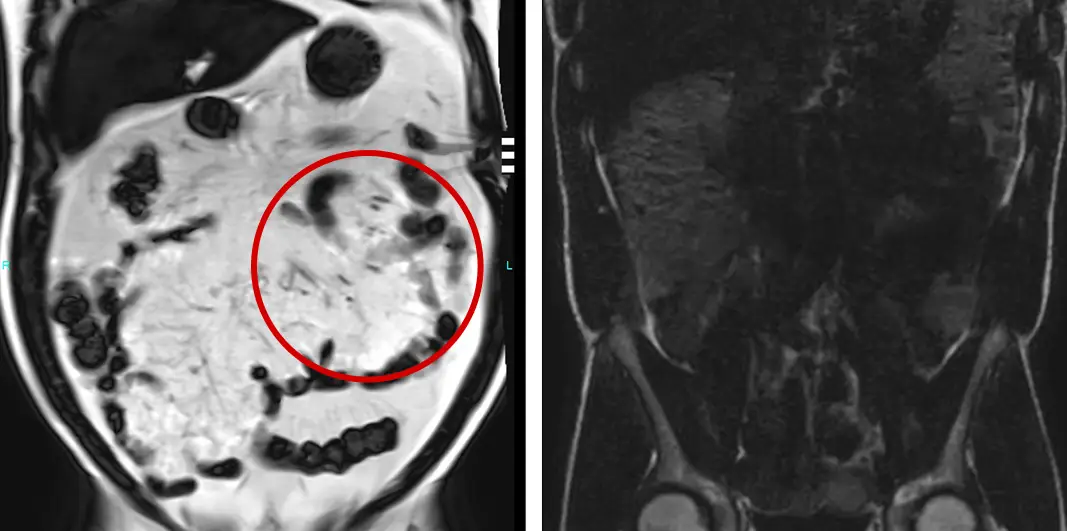
Coronal T1 fat only sequence demonstrates increased visceral fat [Left Image; Red Circle] compared to a patient with a more metabolically-healthy body composition profile with low visceral fat (Right Image).
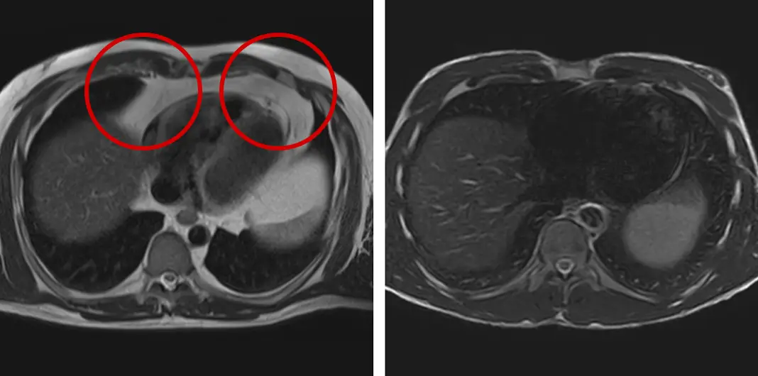
Axial T2-weighted sequence demonstrates increased pericardial fat (Left Image; Red Circles) compared to another patient with more metabolically-healthy body composition profile with low pericardial fat (Right Image).
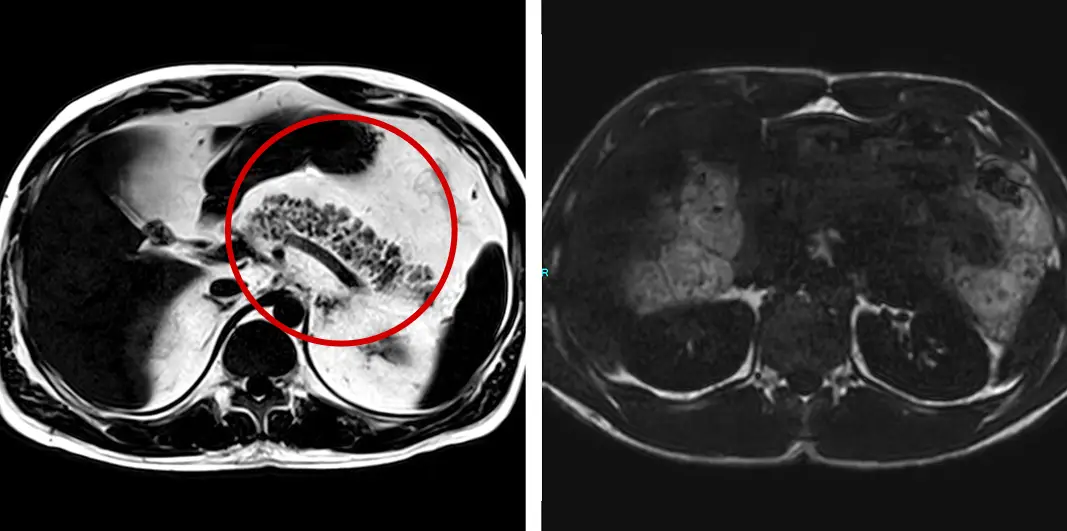
Axial T1 fat only sequence demonstrates increased pancreatic fat (Left Image; Red Circle) compared to another patient with a more metabolically-healthy body composition profile with no abnormally increased pancreatic fat (Right Image).
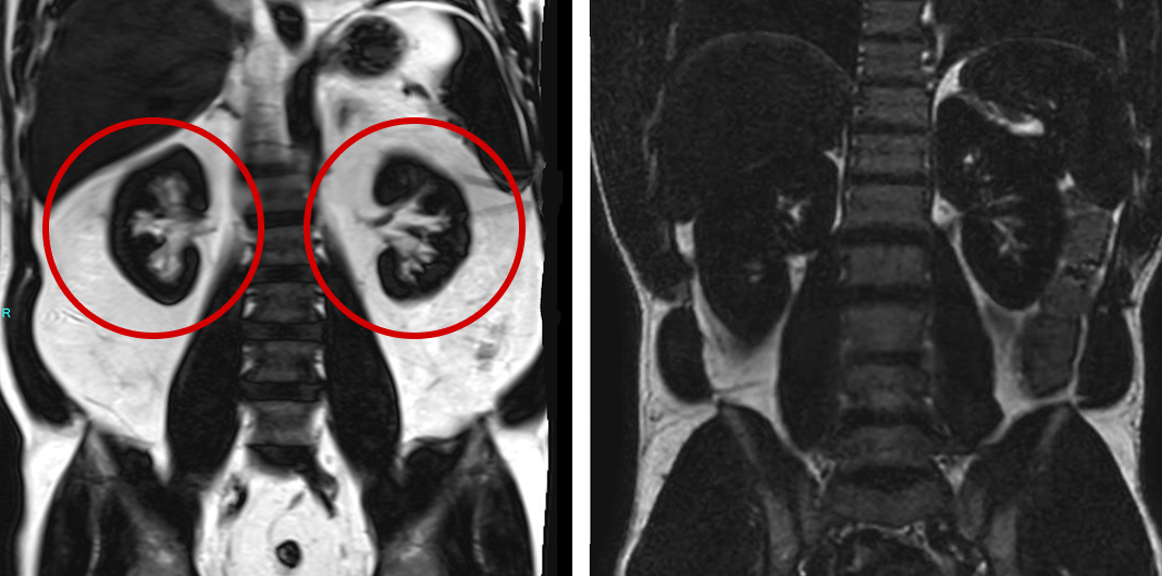
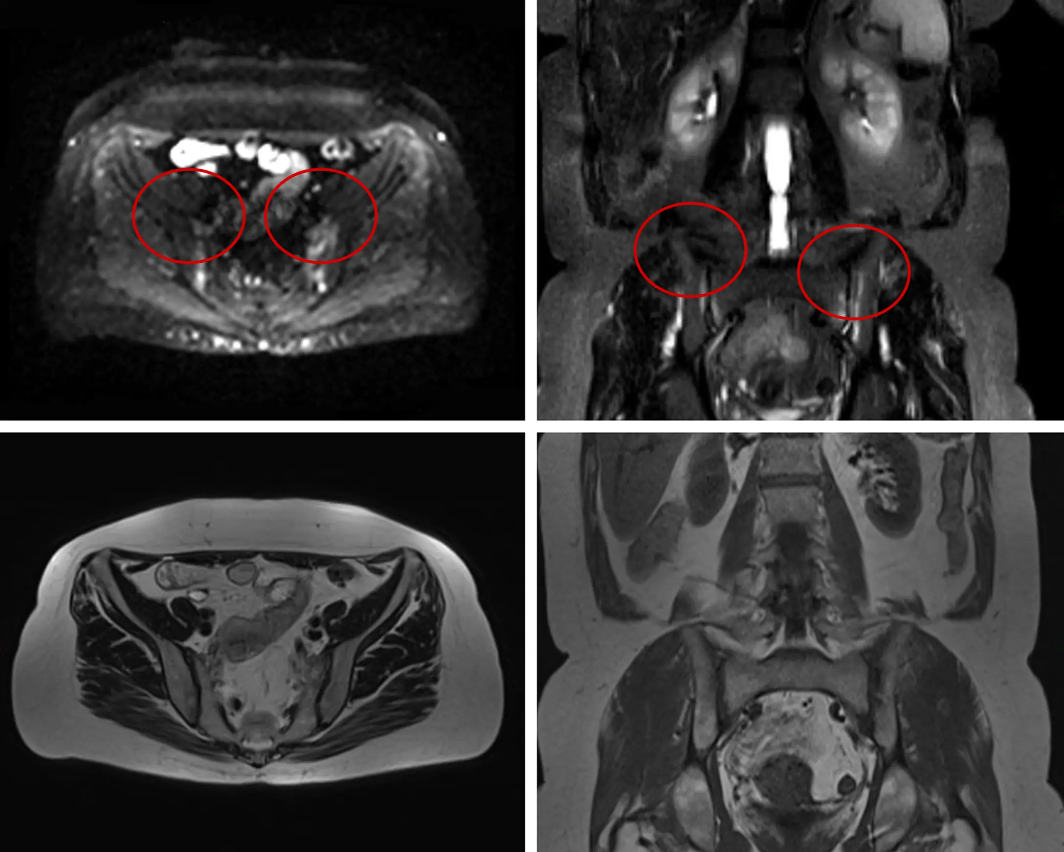
Coronal T1 fat only sequence demonstrates increased renal sinus fat (Left Image; Red Circles) compared to another patient with a more metabolically-healthy body composition profile with low renal sinus fat (Right Image).
Body composition analysis demonstrates greater than 95th percentile visceral fat normalized for age and height.
Coronal T1 fat only sequence demonstrates increased visceral fat [Left Image; Red Circle] compared to a patient with a more metabolically-healthy body composition profile with low visceral fat (Right Image).
Axial T2-weighted sequence demonstrates increased pericardial fat (Left Image; Red Circles) compared to another patient with more metabolically-healthy body composition profile with low pericardial fat (Right Image).
Axial T1 fat only sequence demonstrates increased pancreatic fat (Left Image; Red Circle) compared to another patient with a more metabolically-healthy body composition profile with no abnormally increased pancreatic fat (Right Image).
Coronal T1 fat only sequence demonstrates increased renal sinus fat (Left Image; Red Circles) compared to another patient with a more metabolically-healthy body composition profile with low renal sinus fat (Right Image).
Follow-up care
The patient’s findings placed him at significantly elevated risk for insulin resistance, cardiovascular disease, and metabolic dysfunction. The recommendation for proactive management and preventive care strategy included targeted dietary changes, exercise with a focus on resistance and aerobic training, stress management, and ongoing metabolic monitoring.
How the Prenuvo scan impacted patient care:
The scan results shifted his view of his health—he went from “feeling fine” to realizing he was at risk for future complications. Comparing his scan to that of a healthier, similarly-aged male with significantly less visceral and subcutaneous fat provided a powerful visual contrast, reinforcing the urgency to take action while change is still possible.
References
Other case studies
Proactive WB-MRI Detects Sacroiliitis, A Key Indicator of Inflammatory Arthritis

Subdural Hematoma
